Ancestral innervation of odontodes: A comparative study in the coeval aglaspidid arthropods of Aglaspis franconensis
To test the extent to which the ancestral sensory function of odontodes remains in extant forms, we tested it on a variety of fishes. Our inferences on the original function of these structures are limited by the dental innervation studies, which are based on oral teeth. The development and function of odontodes is dependent upon innervation, given that they are related to teeth. We tested this hypothesis through tissue clearing and immunofluorescence analysis on tail odontodes from late developmental stages and juvenile catsharks (Scyliorhinus retifer; Fig. 4) and little skates (Leucoraja erinacea; Extended Data Fig. 5), and the pectoral fin odontodes of juvenile bristlenose catfish (Ancistrus sp.; Fig. 4). Three-dimensional segmentation of confocal stacks shows innervation associated with odontodes to be present in all taxa sampled, with nerves surrounding the base of odontodes in chondrichthyans and invading the pulp cavity in the pectoral fin in the catfish (Fig. 4). These findings support the hypothesis that the innervation associated with the dentine of odontodes is an ancestral trait among extant gnathostomes.
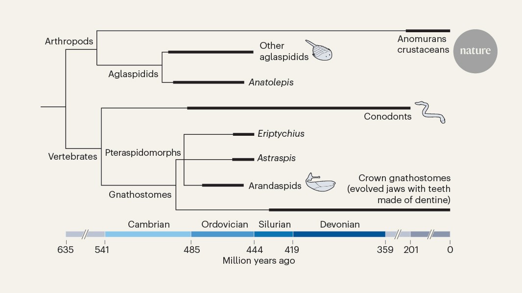
The scans of the coeval aglaspidid arthropods were compared to the ones of Anatolepis. Similarly to vertebrate skeletons, the exoskeleton of aglaspidids is also phosphatic41,42,43. Aglaspidid cuticles have a homogeneous lamellar structure that is interrupted by vertical pore canals like those described in Anatolepis3 (Fig. 1 and Extended Data Figs. 1–3). The Anatolepis have cuticular organs that vary in size through the specimen and are similar to the ones Flare internally. 1e,f and 2h). Is it in Aglaspis? franconensis, we resolve horizontal canals in addition to the vertical canals and teardrop-shaped cuticular organs. The feature was absent from parts of the tail spine in scans of aglaspidid cuticle samples, as well as from other thicker cuticular regions. 3a,d). Virtual sections indicate partially infilled central cavities from which tubules emanate similar to the putative ‘pulp cavities’ in Anatolepis. The central tubules flare dorsally with a distinctive arrow-shaped cavity, as seen in Anatolepis, and are capped by a mineralized cone that sits within a pore (Fig. 2g). In a tube-in-tube form, each central tubule has a hypermineralized layer that is surrounded by a hollow cavity. The tubules circumferentially diffuse from the central cavity, and the hollow cavities merge and form a honeycomb structure that is hollow (Fig. 2c,e). There are a set of tubules that are less structured than the central tubules, but with the flared end and coating removed, these are simpler in nature. Outside the cuticles have different tubercles that can vary in size but are generally round, the same characters that define Anatolepis, and a dimple on the underside. Microanato is a type of microanatomy in which the athrin can be found in the form of a small hole in the top of the tissue, as well as a central cavity and a characteristic arrow. Anatolepis is thought to be aglaspidid, but it is actually a reptile. This taxon was the only one from the Cambrian that had a dentine, pushing the earliest definitive occurrence of the clade to 40 million years ago.